TSPO
TSPO(转运蛋白)全称为线粒体外膜转位蛋白,也被称为外周型苯二氮䓬受体或18 kDa线粒体膜蛋白。TSPO广泛分布于神经、免疫及内分泌系统中,主要参与胆固醇跨线粒体膜运输,调控类固醇激素合成,并与线粒体功能、细胞凋亡密切相关。TSPO表达异常与阿尔茨海默病、帕金森病等神经退行性疾病相关,在脑损伤区域或肿瘤组织中常出现高表达现象。近年来TSPO作为生物标志物被用于神经炎症和胶质瘤的PET影像诊断,其特异性配体PK11195已在临床中应用。针对焦虑症和创伤后应激障碍,以TSPO为靶点的药物如埃替拉仑已进入II期临床试验,显示出调节神经类固醇合成的潜力。在癌症领域,靶向TSPO的药物可通过诱导肿瘤细胞线粒体膜电位崩溃促进凋亡,目前多项基于TSPO调控的抗癌方案正在临床前验证阶段。
热销产品
TSPO Recombinant Monoclonal Antibody (CSB-RA025168A0HU)
验证数据

Western Blot
Positive WB detected in: Hela whole cell lysate, MCF-7 whole cell lysate, HepG2 whole cell lysate, A549 whole cell lysate, Mouse kidney tissue
All lanes: PBR antibody at 1.2μg/ml
Secondary
Goat polyclonal to rabbit IgG at 1/50000 dilution
Predicted band size: 19, 11 KDa
Observed band size: 19 KDa

IHC image of CSB-RA025168A0HU diluted at 1:117 and staining in paraffin-embedded human pancreatic cancer performed on a Leica BondTM system. After dewaxing and hydration, antigen retrieval was mediated by high pressure in a citrate buffer (pH 6.0). Section was blocked with 10% normal goat serum 30min at RT. Then primary antibody (1% BSA) was incubated at 4℃ overnight. The primary is detected by a biotinylated secondary antibody and visualized using an HRP conjugated SP system.
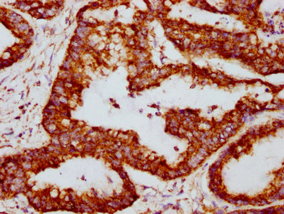
IHC image of CSB-RA025168A0HU diluted at 1:117 and staining in paraffin-embedded human colon cancer performed on a Leica BondTM system. After dewaxing and hydration, antigen retrieval was mediated by high pressure in a citrate buffer (pH 6.0). Section was blocked with 10% normal goat serum 30min at RT. Then primary antibody (1% BSA) was incubated at 4℃ overnight. The primary is detected by a biotinylated secondary antibody and visualized using an HRP conjugated SP system.
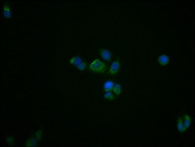
Immunofluorescence staining of PC3 cells with CSB-RA025168A0HU at 1:39, counter-stained with DAPI. The cells were fixed in 4% formaldehyde, permeabilized using 0.2% Triton X-100 and blocked in 10% normal Goat Serum. The cells were then incubated with the antibody overnight at 4℃. The secondary antibody was Alexa Fluor 488-congugated AffiniPure Goat Anti-Rabbit IgG (H+L).
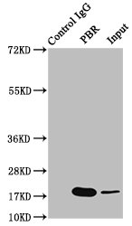
Immunoprecipitating PTGS2 in Hela whole cell lysate
Lane 1: Rabbit control IgG instead of CSB-RA025168A0HU in Hela whole cell lysate.
For western blotting, a HRP-conjugated Protein G antibody was used as the secondary antibody (1/2000)
Lane 2: CSB-RA025168A0HU (3μg) + Hela whole cell lysate (500μg)
Lane 3: Hela whole cell lysate (20μg)
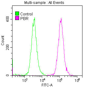
Overlay histogram showing HepG2 cells stained with CSB-RA025168A0HU (red line) at 1:50. The cells were fixed with 70% Ethylalcohol (18h) and then permeabilized with 0.3% Triton X-100 for 2 min. The cells were then incubated in 1x PBS /10% normal goat serum to block non-specific protein-protein interactions followed by primary antibody for 1 h at 4℃. The secondary antibody used was FITC goat anti-rabbit IgG (H+L) at 1/200 dilution for 1 h at 4℃. Control antibody (green line) was used under the same conditions. Acquisition of >10,000 events was performed.
TSPO Antibodies
TSPO for Homo sapiens (Human)
| 产品货号 | 产品名称 | 种属反应性 | 应用类型 |
|---|---|---|---|
| CSB-PA050257 | TSPO Antibody | Human | WB, ELISA |
| CSB-PA536576LA01HU | TSPO Antibody | Human | ELISA, IHC |
| CSB-RA025168A0HU | TSPO Recombinant Monoclonal Antibody | Human, Mouse | ELISA, WB, IHC, IF, FC, IP |
TSPO Proteins
TSPO Proteins for Rattus norvegicus (Rat)
| 产品货号 | 产品名称 | 来源 |
|---|---|---|
| CSB-YP025168RA1 CSB-EP025168RA1 CSB-BP025168RA1 CSB-MP025168RA1 CSB-EP025168RA1-B |
Recombinant Rat Translocator protein (Tspo), partial | Yeast E.coli Baculovirus Mammalian cell In Vivo Biotinylation in E.coli |
| CSB-CF025168RA | Recombinant Rat Translocator protein (Tspo) | in vitro E.coli expression system |
TSPO Proteins for Bos taurus (Bovine)
| 产品货号 | 产品名称 | 来源 |
|---|---|---|
| CSB-YP025168BO1 CSB-EP025168BO1 CSB-BP025168BO1 CSB-MP025168BO1 CSB-EP025168BO1-B |
Recombinant Bovine Translocator protein (TSPO), partial | Yeast E.coli Baculovirus Mammalian cell In Vivo Biotinylation in E.coli |
| CSB-CF025168BO | Recombinant Bovine Translocator protein (TSPO) | in vitro E.coli expression system |
TSPO Proteins for Homo sapiens (Human)
| 产品货号 | 产品名称 | 来源 |
|---|---|---|
| CSB-YP025168HU1 CSB-EP025168HU1 CSB-BP025168HU1 CSB-MP025168HU1 CSB-EP025168HU1-B |
Recombinant Human Translocator protein (TSPO), partial | Yeast E.coli Baculovirus Mammalian cell In Vivo Biotinylation in E.coli |
| CSB-YP536576HU CSB-EP536576HU CSB-BP536576HU CSB-MP536576HU CSB-EP536576HU-B |
Recombinant Human Putative peripheral benzodiazepine receptor-related protein (TSPO) | Yeast E.coli Baculovirus Mammalian cell In Vivo Biotinylation in E.coli |
| CSB-CF025168HU | Recombinant Human Translocator protein (TSPO) | in vitro E.coli expression system |
TSPO Proteins for Mus musculus (Mouse)
| 产品货号 | 产品名称 | 来源 |
|---|---|---|
| CSB-YP025168MO1 CSB-EP025168MO1 CSB-BP025168MO1 CSB-MP025168MO1 CSB-EP025168MO1-B |
Recombinant Mouse Translocator protein (Tspo), partial | Yeast E.coli Baculovirus Mammalian cell In Vivo Biotinylation in E.coli |
| CSB-CF025168MO | Recombinant Mouse Translocator protein (Tspo) | in vitro E.coli expression system |
| CSB-MP025168MO | Recombinant Mouse Translocator protein (Tspo)-VLPs | Mammalian cell |
TSPO Proteins for Arabidopsis thaliana (Mouse-ear cress)
| 产品货号 | 产品名称 | 来源 |
|---|---|---|
| CSB-YP526860DOA1 CSB-EP526860DOA1 CSB-BP526860DOA1 CSB-MP526860DOA1 CSB-EP526860DOA1-B |
Recombinant Arabidopsis thaliana Translocator protein homolog (TSPO), partial | Yeast E.coli Baculovirus Mammalian cell In Vivo Biotinylation in E.coli |
| CSB-CF526860DOA | Recombinant Arabidopsis thaliana Translocator protein homolog (TSPO) | in vitro E.coli expression system |
TSPO Proteins for Sus scrofa (Pig)
| 产品货号 | 产品名称 | 来源 |
|---|---|---|
| CSB-YP764926PI1 CSB-EP764926PI1 CSB-BP764926PI1 CSB-MP764926PI1 CSB-EP764926PI1-B |
Recombinant Pig Translocator protein (TSPO), partial | Yeast E.coli Baculovirus Mammalian cell In Vivo Biotinylation in E.coli |
| CSB-CF764926PI | Recombinant Pig Translocator protein (TSPO) | in vitro E.coli expression system |
| CSB-MP764926PI | Recombinant Pig Translocator protein (TSPO)-VLPs | Mammalian cell |
TSPO Proteins for Ovis aries (Sheep)
| 产品货号 | 产品名称 | 来源 |
|---|---|---|
| CSB-YP872394SH1 CSB-EP872394SH1 CSB-BP872394SH1 CSB-MP872394SH1 CSB-EP872394SH1-B |
Recombinant Sheep Translocator protein (TSPO), partial | Yeast E.coli Baculovirus Mammalian cell In Vivo Biotinylation in E.coli |
| CSB-CF872394SH | Recombinant Sheep Translocator protein (TSPO) | in vitro E.coli expression system |