PDCD1
PDCD1,即程序性细胞死亡蛋白1,也称为PD1、CD279、hSLE1,由人类PDCD1基因编码,该基因位于2号染色体的q37.3位置。PDCD1蛋白是一种重要的免疫抑制受体,属于免疫球蛋白超家族成员,主要在激活的T细胞和B细胞上表达
其作用机制涉及与配体PD-L1和PD-L2的结合,启动T细胞的程序性死亡,从而抑制T细胞的活化和增殖。
PDCD1在维持外周T、B细胞耐受中发挥重要作用,其增强信号可以下调已活化的T、B细胞在外周组织中的应答。PDCD1的异常表达与多种自身免疫疾病、感染性疾病和肿瘤相关,是免疫治疗的重要靶点。
热销产品
Recombinant Human Programmed cell death protein 1 (PDCD1), partial (Active) (CSB-MP619964HU1)
验证数据
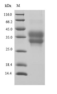
(Tris-Glycine gel) Discontinuous SDS-PAGE (reduced) with 5% enrichment gel and 15% separation gel.
Activity
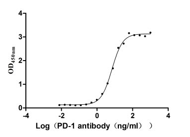
①Measured by its binding ability in a functional ELISA. Immobilized PD-1 at 2 μg/ml can bind Anti-PD-1 recombinant antibody, the EC50 of human PD-1 protein is 6.087-7.854 ng/ml.
Activity
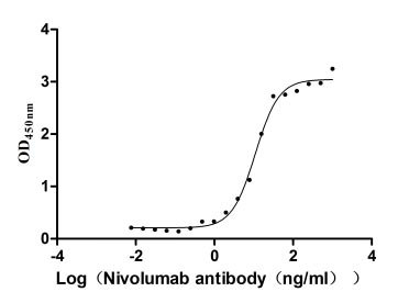
②Measured by its binding ability in a functional ELISA. Immobilized PD-1 at 2 μg/ml can bind Nivolumab, the EC50 of human PD-1 protein is 9.713-12.39 ng/ml.
PDCD1 Recombinant Monoclonal Antibody (CSB-RA240597A0HU)
验证数据
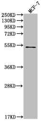
Western Blot
Positive WB detected in: MCF-7 whole cell lysate
All lanes: PD1 antibody at 1:2000
Secondary
Goat polyclonal to rabbit IgG at 1/50000 dilution
Predicted band size: 32 KDa
Observed band size: 32 kDa
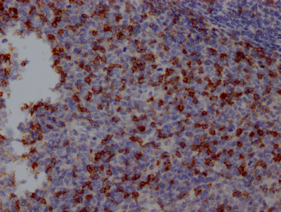
IHC image of CSB-RA240597A0HU diluted at 1:100 and staining in paraffin-embedded human tonsil tissue performed on a Leica BondTM system. After dewaxing and hydration, antigen retrieval was mediated by high pressure in a citrate buffer (pH 6.0). Section was blocked with 10% normal goat serum 30min at RT. Then primary antibody (1% BSA) was incubated at 4℃ overnight. The primary is detected by a Goat anti-rabbit IgG polymer labeled by HRP and visualized using 0.05% DAB.
PDCD1 Antibodies
PDCD1 for Homo sapiens (Human)
| 产品货号 | 产品名称 | 种属反应性 | 应用类型 |
|---|---|---|---|
| CSB-PA017664GA01HU | PDCD1 Antibody | Human | ELISA,WB,IHC |
| CSB-PA483633 | PDCD1 Antibody | Human | ELISA,IHC |
| CSB-PA619964LA01HU | PDCD1 Antibody | Human, Mouse | ELISA, WB, IHC, IF |
| CSB-PA619964LD01HU | PDCD1 Antibody, Biotin conjugated | Human | ELISA |
| CSB-PA619964LC01HU | PDCD1 Antibody, FITC conjugated | Human | |
| CSB-PA619964LB01HU | PDCD1 Antibody, HRP conjugated | Human | ELISA |
| CSB-RA240597A0HU | PDCD1 Recombinant Monoclonal Antibody | Human | ELISA, WB, IHC |
| CSB-MA619964A0m | PD-1 Monoclonal Antibody | Human | ELISA, WB |
| CSB-MA619964A1m | PD-1 Monoclonal Antibody | Human | ELISA, WB |
| CSB-RA619964MA1HU | PD-1 Recombinant Monoclonal Antibody | Human | ELISA |
PDCD1 Proteins
PDCD1 Proteins for Homo sapiens (Human)
| 产品货号 | 产品名称 | 来源 |
|---|---|---|
| CSB-YP619964HU CSB-BP619964HU CSB-MP619964HU CSB-EP619964HU-B |
Recombinant Human Programmed cell death protein 1 (PDCD1), partial | Yeast Baculovirus Mammalian cell In Vivo Biotinylation in E.coli |
| CSB-EP619964HU | Recombinant Human Programmed cell death protein 1 (PDCD1), partial | E.coli |
| CSB-MP619964HU1 | Recombinant Human Programmed cell death protein 1 (PDCD1), partial (Active) | Mammalian cell |
PDCD1 ELISA Kit
PDCD1 ELISA Kit for Homo sapiens (Human)
| 产品货号 | 产品名称 | 样本类型 | 灵敏度 |
|---|---|---|---|
| CSB-E13643h | Human Programmed Death 1(PD-1)ELISA KIT | serum, plasma, tissue homogenates, cell lysates | 3.9 pg/mL |
PDCD1 ELISA Kit for Mus musculus (Mouse)
| 产品货号 | 产品名称 | 样本类型 | 灵敏度 |
|---|---|---|---|
| CSB-E13586m | Mouse Programmed Death 1(PD-1)ELISA Kit | serum, plasma, tissue homogenates, cell lysates | 0.078 ng/mL |